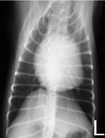
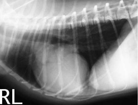
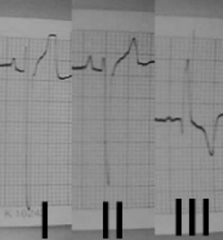
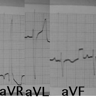
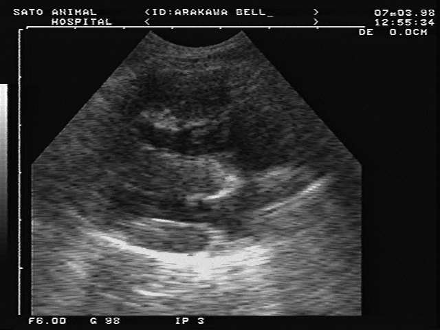
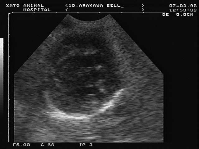
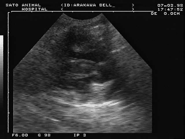
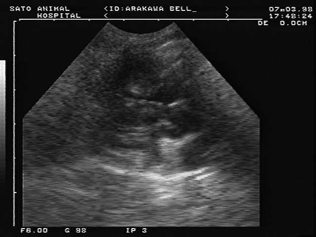
No.2(Pink)TOF?
A 2-year-old female Chihuahua(2.1kg) with III/VI systoric murmur (MPI in the pulmonic valuve region).Å@Radiographs shows enlargement of heart size, inverted D-shape of the heart and diminished pulmonary vasculature, increased contact between the right ventricular and sternum(VD and RL views).Å@Electrocardiogram shows right ventricular hypertrophy pattern.Å@Echocardiography reveals right ventricular hypertrophy, thickened ventricular septum.Å@Bubble study shows right-to-aorta shuntning. Mild polycythemia (RBC 709, PCV 50.3, Hb 15.3, MCV 71, MCH 21.6, MCHC30.4) is seen.Å@Hx of demodicosis. Just one episode of syncope. No cyanosis. Å@
No.3 Chronic bronchitis?(English)
No.4 DCM?(English)
Sato Animal Hospital
tsato@usa.net
tsato@usa.net